



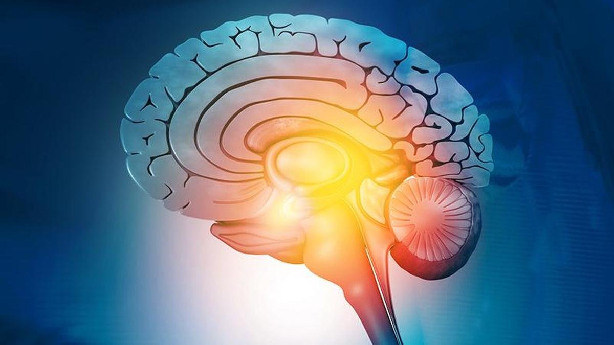
A study published in September in Nature by neuroscientist Sergiu Pașca’s group at Stanford University reports an experiment that sounds almost like science fiction. Human brain tissue grown from stem cells was transplanted into mice in which much of the cerebral cortex had been genetically prevented from forming. The transplanted tissue grew into this vacant space, produced several types of human brain cells, became electrically active, connected with the mouse nervous system and was associated with measurable differences in behaviour.
Yet, an important limitation must be stated at the outset. The researchers have not created a human brain inside a mouse. The transplanted tissue remained immature, lacked the ordered structure of a normal human cortex, and the study cannot establish that human neurons directly caused any particular behaviour. What the experiment offers is a narrow glimpse of human brain development as it unfolds inside a non-human living nervous system.
Our blind spot
Understanding how the human brain develops matters both for evolutionary biology and for medicine. The cerebral cortex expanded greatly during human evolution, developing specialised cells and connections involved in language, planning and complex social behaviour. Studying how these features emerge may help explain what makes the human brain distinctive.
It is also medically important because many neurological and psychiatric disorders may begin during development, long before symptoms appear. The main challenge is access. Scientists cannot experimentally manipulate a developing human brain as they can in laboratory animals. Imaging can track structural changes, while foetal and post-mortem tissues provide useful snapshots. Still, neither allows researchers to alter a gene or developmental process and observe the consequences over time. Human developmental neuroscience has therefore long faced a major experimental blind spot.
Growing brain tissue
Stem cell technology opened up another avenue to study the developing human brain. Scientists can take ordinary human cells, re-programme them into an earlier, flexible state called induced pluripotent stem cells, and guide them to become nerve cells. When these cells are grown together in three dimensions, they can organise into structures called organoids. A brain organoid is a small piece of laboratory-grown neural tissue that reproduces some features of early brain development. Organoids allow researchers to observe how human neurons develop, introduce disease-related genetic changes and test responses to drugs.
However, tissue grown in a lab dish lacks key features of a living brain, including normal blood circulation, sensory inputs and long-distance connections with other parts of the nervous system. Researchers, including the Stanford group, have therefore transplanted human brain organoids into rodents, where the living environment supports better survival and maturation. Yet this creates another constraint: the rodent already has its own cortex, so the transplanted human tissue must compete for limited space and neural connections, and has never amounted to more than a fragment of the host’s brain.
A third window
The Stanford researchers opened up a third possibility. Instead of engineering a better graft, they re-engineered the host. Using genetic techniques, they produced mice in which much of the neocortex and hippocampus never formed. Because the mice were also bred to lack a functioning immune system, the human tissue would not be rejected. Human cortical organoids were then transplanted into the vacant space soon after birth. The researchers call the approach xenocortication. ‘Xeno’ refers to something originating from another species, while the cortex is the outer region of the brain involved in many higher functions.
In the Stanford experiment, freed from much of the competition from mouse cortical tissue, the human grafts expanded substantially, and by three months, they made up about 92% of the animal’s cortical tissue. They produced several types of developing cortical neurons, along with progenitor cells and astrocytes, at a stage of maturity roughly equivalent to that of the human cortex late in the second trimester of pregnancy. The grafts generated deep-layer neurons carrying the molecular signature of von Economo neurons, large, distinctively shaped cells found in humans and a few other large-brained mammals, but absent in rodents, and long associated with social cognition and psychiatric illness. Such neurons are almost impossible to grow in a dish.
The grafts also became electrically active, producing slow waves that swept across the tissue every few minutes, a pattern characteristic of developing, rather than mature, cortical networks. They formed connections with surviving parts of the mouse nervous system, and human nerve fibres reached as far as the cervical spinal cord, something not seen in mice with an intact cortex. This is the conceptual leap of the study: human neural tissue was no longer a fragment lodged inside a rodent’s brain, but tissue occupying almost all of the space where that animal’s cortex would have been.
Reading behaviour
The next question was whether this integration had any functional effect. The starting point was itself surprising. Mice lacking most of their neocortex and hippocampus (roughly half the brain by volume) remained active and mobile, ran at normal speeds and responded normally to heat and touch. To detect subtler differences, the researchers combined conventional behavioural tests with a machine-learning system called Motion Sequencing (MoSeq). A depth camera recorded freely moving mice, while the software divided their movements into 60 recurring sub-second patterns, such as rearing, pausing, and turning, creating a detailed behavioural fingerprint rather than relying solely on speed or distance travelled. By this measure, mice lacking much of their cortex behaved differently from normal animals. Xenocortical mice showed an intermediate pattern, suggesting that transplantation altered behaviour without fully restoring it. In a Y-shaped maze used to test working memory, normal and xenocortical mice performed above chance, whereas mice lacking much of their cortex did not. This suggests possible partial functional compensation, but does not prove that human neurons restored memory.
Why this matters
To show what the platform can do, the researchers exposed the mice to five hours of severe oxygen deprivation. The response was confined to the human tissue: a molecular marker of oxygen stress appeared in the graft but not in the adjacent mouse brain, and not at all in animals without a graft. Two days later, the xenocortical mice walked differently, shifting to a more stable stance without any change in their speed. Imaging 10 days on showed changes in the graft’s blood vessels. Under the microscope, the graft’s supporting cells had multiplied and become more complex.
This creates a potentially powerful experimental pathway. Scientists could introduce a disease-related mutation into human cells, allow the resulting neural tissue to develop within a living nervous system, observe changes in cells and circuits, assess whether behaviour is affected, and then test possible treatments. Such models could help study developmental brain disorders, the oxygen deprivation around birth that can lead to cerebral palsy, human-specific neuronal diseases and evolutionary differences between human and animal brains. They may also provide a platform for drug testing and, eventually, for evaluating regenerative or cell-based treatments for severe early brain injury.
Limits and ethics
The model is still far from reproducing a developing human brain. The grafts did not reproduce the full balance of excitatory and inhibitory neurons. Human neurons also mature far more slowly than mouse neurons, creating a mismatch between the developmental timelines of graft and host. Although anatomical connections were observed, this does not prove that they directly controlled behaviour. Any behavioural changes, therefore, arise from a hybrid system in which human tissue interacts with the remaining mouse brain.
These scientific limits also shape the ethical questions. There is no evidence that the animals developed human consciousness or a human-like mind. However, future models may contain larger, more mature, and better-organised human neural tissue, with greater integration into the host brain. At what point might this alter an animal’s cognition, perception or capacity for experience in ethically significant ways? The researchers anticipated this question and consulted bioethicists at Stanford before beginning. The paper argues that going further towards grafts with mature cortical layering, or transplantation into embryos or into non-human primates, will require a greater degree of scrutiny before such experiments begin.
Ultimately, xenocortication is important not because it creates a human brain in another species, but because it may provide a third window to study human brain development.
(Dr. C. Aravinda is an academic and public health physician. aravindaaiimsjr10@hotmail.com; Dr Veera Rajagopal is a physician-scientist & geneticist working in drug discovery. veera.scientist@gmail.com)
Published – October 02, 2026 06:00 am IST
