



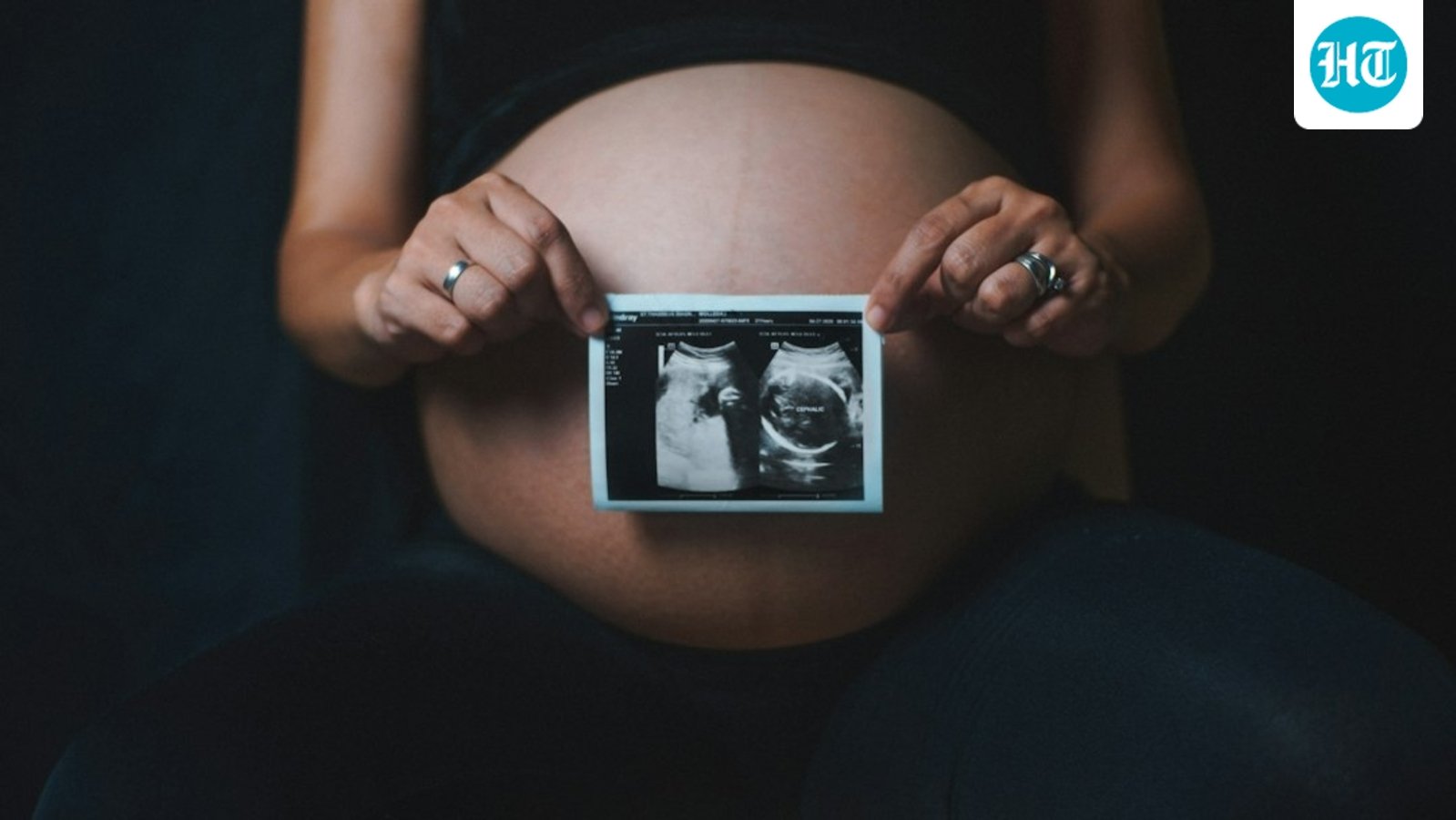
Finding out you are pregnant can suddenly make even routine medical decisions feel more complicated. Something as familiar as an X-ray may prompt questions about whether radiation could affect the pregnancy or the developing baby. These concerns are understandable, but pregnancy does not automatically mean that every X-ray or imaging examination must be avoided.
Also Read | Are X-ray scans really harmful? Radiologist debunks 5 health-related myths fuelled by social media
Whether an investigation is recommended depends on several factors, including the type of examination, the part of the body being examined, the amount of radiation involved and, importantly, whether the test is necessary for the mother’s health. When imaging is medically indicated, doctors weigh its potential risks against the benefits of getting an accurate and timely diagnosis.
In an interview with HT Lifestyle, Dr Bimalpreet Mohan, the director, chief radiologist and women’s health expert at Capital Health Clinic in Hauz Khas, New Delhi, explains, “The first thing I tell pregnant women is not to panic if an X-ray has been recommended. They should tell the doctor and the radiology team that they are pregnant or may be pregnant. Once we know that, we can consider the safest and most appropriate way to investigate the problem.”
Why the location of the X-ray matters
According to Dr Mohan, not all X-rays expose the developing baby to the same amount of radiation. An examination of the chest, arm, leg or head, for example, is very different from imaging that directly involves the abdomen or pelvis.
For abdominal or pelvic examinations, the pregnancy and the medical necessity of the test need to be considered carefully. Doctors may explore whether another appropriate imaging method can provide the required information. Ultrasound, which does not use ionising radiation, is commonly used during pregnancy, while MRI may also be appropriate in selected circumstances.
“There is no single rule that says an X-ray can never be performed during pregnancy. We look at what needs to be diagnosed and whether the information can be obtained through another suitable investigation. If an X-ray is genuinely required, the examination is planned with radiation protection in mind,” highlights Dr Mohan.
What if the woman is unaware of the pregnancy?
Some women may undergo an X-ray before discovering that they are pregnant. A woman in this situation should inform her doctor rather than immediately assuming that harm has occurred. The actual exposure depends on the examination and the area of the body imaged, so the circumstances need to be reviewed individually.
The radiologist points out, “An accidental X-ray before a woman knew she was pregnant should not automatically become a reason for panic. We need to know what examination was performed and when. The radiology team can assess the exposure and guide the patient appropriately.”
Can shielding make the examination safer?
Radiation protection is an important part of radiology. Depending on the examination and local protocols, appropriate positioning, equipment settings and protective measures may be used to minimise unnecessary exposure.
Dr Mohan highlights that the larger principle is justification and optimisation: an examination should have a clear medical reason, and radiation exposure should be kept as low as reasonably achievable while still obtaining useful diagnostic information.
Should women choose an ultrasound instead?
Ultrasound is extremely useful during pregnancy, but it cannot answer every clinical question that an X-ray or another imaging examination can. The radiologist explains, “Patients sometimes think that because ultrasound has no ionising radiation, it can replace every other imaging test. That is not the case. Each modality gives us different information. The right choice depends on what we are looking for and which test can answer that particular clinical question.”
Pregnancy should make women more informed about imaging, not fearful of it. Always tell your doctor and radiologist about a confirmed or possible pregnancy before an examination. Ask why the scan is required, whether another suitable test could provide the same information and how the examination will be performed safely.
Dr Mohan concludes, “The aim is never to expose a pregnant woman unnecessarily. But delaying an important diagnosis can also carry risks. Imaging decisions during pregnancy should therefore be individualised, with the health of both mother and baby kept in mind.”
Note to readers: This article is for informational purposes only and not a substitute for professional medical advice. Always seek the advice of your doctor with any questions about a medical condition.
Dr Bimalpreet Mohan is the director and head of the department of radiology at Capital Health & Diagnostic Clinic, Hauz Khas, New Delhi, with over 15 years of experience in advanced diagnostic imaging. She specialises in MRI, CT scans, ultrasound, doppler imaging, mammography and breast imaging.
